
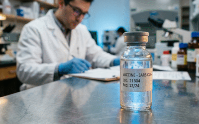
Dr. Philip McMillan, John McMillan
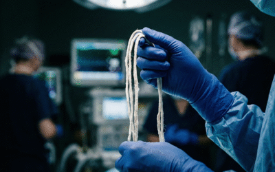
Something changed in 2021. Embalmers, the professionals who drain blood from the deceased and replace it with preservation fluid, started pulling out things they had never seen. Long, white, rubbery structures, some stretching 25 centimeters, clogging arterial and venous lines. Not soft, crumbly red clots but elastic, fibrous casts that resisted every attempt at breakdown.
U.S. embalmer Richard Hirschman was among the first to go public. One specimen filled a 19-inch tube. A global survey by Tom Haviland found 83% of embalmers encountering these white structures since 2021, with up to 27.5% of bodies containing them. A recent webinar brought together three researchers working to make sense of it: Philip McMillan, a clinician-researcher tracking post-COVID molecular biology; Bruce Rafley, a New Zealand-based applied biologist; and Dr. Matt Shelton, a doctor of 38 years involved with NZDSOS, New Zealand’s medical freedom movement.
The Blackout: Why Conventional Science Couldn’t Respond
If these clots are real (and the preprint papers now emerging from Rafley and Shelton’s team confirm that they are) why hasn’t the scientific establishment already figured this out? The answer, according to Rafley, is blunt. Laboratories around the world were explicitly told by government agencies that they could not investigate anything related to COVID-19. Not discouraged. Forbidden.
“When I tried to dig deeper into this and say ‘Who has told you this?’ everything went quiet,” Rafley recounted. “Our policy direction: we’ve been directed not to work on COVID.” University labs, private facilities, institutional researchers: all blocked from lines of inquiry outside the approved narrative. Autopsies on individuals who had supposedly died of COVID were almost impossible to obtain. The normal scientific reflex in a health emergency was nowhere to be found.
Rafley’s workaround was born of necessity. He contacted embalmers from three countries, secured samples, and submitted them on a black-box basis to international laboratories: no names, no affiliations, no context. It was the only way to access specialized equipment without triggering institutional gatekeeping. The result is a set of three preprint papers representing the first serious morphological and proteomic characterization of these structures. The unconventional methodology was not a weakness but a direct consequence of an environment that made conventional research impossible.
Deconstructing the Clots: What the Black-Box Revealed
A normal blood clot is straightforward: fibrin strands trap red blood cells, creating a soft, dark-red mass that enzymes dissolve once bleeding stops. These white structures are something else entirely, nearly devoid of red blood cells, elastic, rubbery, layered, growing progressively over weeks to months, and resistant to the body’s standard dissolution mechanisms.
Rafley’s lab analysis revealed what he called “a complete dog’s breakfast.” Not clean biological architecture but a chaotic tangle of misfolded, amyloid-like proteins with 532 distinct blood proteins trapped in the mesh, alongside broken fibrinogen, fragmented blood cells, and platelet remnants. This is the signature of a system gone haywire, where unsatisfied chemical bonds on damaged fibrin molecules grab onto anything passing through the bloodstream.
Two findings stood out. First, a hemoglobin anomaly: in intact red blood cells, alpha and beta hemoglobin chains exist in a 1:1 ratio. In the white clots, that ratio was skewed to roughly 7:1 in favour of beta chains, meaning something was destroying massive numbers of red blood cells and selectively degrading the less stable alpha chains.
The second finding was the near-total absence of plasminogen, the enzyme the body normally embeds within a clot as a built-in self-destruct mechanism. Rafley put it memorably: “I think of it like the explosive bolts in a rocket. When we send someone to the moon, we need to get rid of the large payload that got them into orbit. So we have explosive bolts that destroy the main engine. In a clot, it’s putting in an enzyme which when activated will take the clot apart.” In these white clots, the explosive bolts are missing. Without plasminogen, and with reduced gamma and alpha fibrin chains for it to bind to even if it were present, the clots simply keep growing. Layer upon layer, week after week.
The Pathophysiological Cascade: How They Form
Shelton offered a key insight about origins. The inflammation, he suggested, starts not on the inner lining of arteries but in the vasa vasorum, the tiny vessels supplying artery walls. If these become blocked, the larger vessels lose oxygen supply. The resulting hypoxia and endothelial damage creates the initial nidus for clot formation, and because the outer inflammation persists, the inner damage never resolves.
Platelets arrive first at the injury site. These sub-cellular fragments, described by Rafley as “little robot cells” running on mRNA programming, detect damage, change shape, extend arms to grip one another, and send chemical distress signals. The liver responds by producing more fibrinogen blindly, which arrives and forms thin protofibrils that attach to the platelet plug like a gossamer fishing net.
Normally, platelets control this process and prevent runaway growth. But here, the process escapes containment. McMillan highlighted a vicious cycle where macrophages release wave after wave of extracellular nets, stimulating neutrophils to do the same. The result is a spider-web trap that indiscriminately captures immunoglobulins, proteins, cell fragments, and the free hemoglobin released from ruptured red blood cells.
At the centre of this cascade sits a cell that McMillan stumbled upon during his research into severe COVID-19: the THBS1 monocyte. This white blood cell appears to drive both hemoglobin breakdown and localized thrombosis simultaneously, a dual role that could explain how hemolysis and clot formation feed each other in a self-reinforcing loop.
Who Is Most Vulnerable?
If hemolysis is the engine driving these clots, then anyone whose red blood cells are already vulnerable occupies higher ground on the risk gradient. McMillan identified glucose-6-phosphate dehydrogenase (G6PD) deficiency, affecting roughly 400 million people worldwide and concentrated in Africa, the Middle East, and India, as a potentially critical factor. G6PD-deficient individuals cannot adequately recycle glutathione, the antioxidant that protects red blood cells from oxidative damage. Under stress, their red cells become fragile and prone to rupture, feeding the hemolysis-clot cycle.
G6PD deficiency is only one piece. McMillan described a “perfect storm” patient: an older individual with metabolic syndrome, autoimmune conditions, and ongoing spike protein circulation. On vaccines, he was direct: repeated mRNA doses trigger a class switch to tolerance-promoting IgG4 antibodies that block viral clearance rather than enabling the immune system. Combined with bypassed mucosal immunity, this drives chronic vascular inflammation. Shelton noted that a New Zealand whistleblower reported during the rollout to the over-65s, roughly 90% of the bodies he embalmed had died within two weeks of injection.
A Practical Defence: Terrain Over Magic Bullets
With conventional medicine not yet acknowledging the problem, McMillan outlined four pillars of natural protection: gut barrier repair to seal the gateway for immune complexes; antioxidant defence with NAC, vitamin C, and selenium; natural fibrinolysis using nattokinase and lumbrokinase; and anti-inflammatory support through omega-3 fatty acids and curcumin. Most people would need only three or four interventions, one from each pillar, at the right dose and time.
Beneath all supplementation sits what Shelton called “profoundly powerful” lifestyle basics: diverse seasonal eating, exercise, quality sleep, and intermittent fasting. The white clot mystery is not fully solved, and more questions than answers remain. But the picture emerging from black-box labs and embalmer’s tables is increasingly coherent: a cascade of hemolysis, runaway fibrin deposition, and missing self-destruct mechanisms affecting far more people than anyone in authority has been willing to count.
This is the scariest thing yet that is being covered up by Govt. It is probably just the tip of the iceberg, among other things that are……..?
I watched your video but reading this is even more eye opening. Thank you.
Thank you for the honest information. I hope and pray doctors will use and understand the information 🙏 to save more lives. In God’s name!
Amen!